Results
|
When stained with methylene blue, the morphological changes in the cells were easier to observe. The projections of the cells, the nuclei, and cell walls were much darker and an easy comparison was made between the RA cells and the Controls as shown in the pictures to the right. The treated cells have dark, thin projections.
|
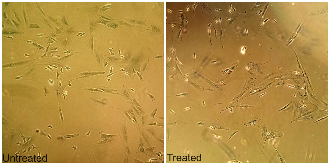
Figure 1 A)
|
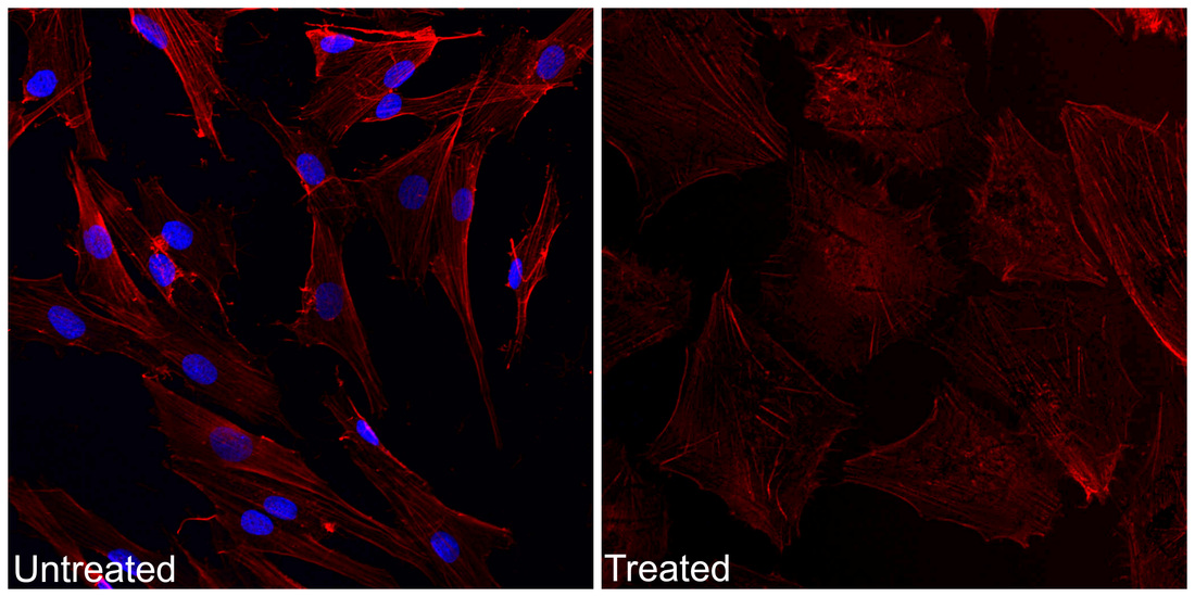
Figure 1 B)
|
Phallodin stained the cell’s skeletal structure. In the picture to the left, the treated cells have defined projections like the methylene blue. Due to error in the staining procedure the dapi blue did not light up under the confocal in the treated cells. The cells treated by RA have defined cells walls.
|
|
The immunofluorescence and antibodies used to stain the cells marked specific proteins in the cell. There are a greater number of cells on the untreated slide since they are at a higher confluency. Green and red markers are in both cells but significantly less red in the untreated cells as well as lower concentration of the green marker. The treated cells have more red stringy projections and defined cells walls.
|
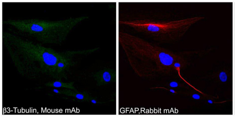
Figure 1 C)
|
Figure 1. Treatment of SK-N-SH cells with Retinoic Acid results in distinct morphological changes. A) Methylene blue staining followed by light microscopy, shown are control DMSO treated and RA treated cells. B) Phalloidin-Fluorescence confocal imaging and C) and Immunofluorescence imaging of the neuronal markers β3-Tubulin (Green) and GFAP (Red). DAPI Nuclear staining in blue was performed for confocal microscopy.
|
|
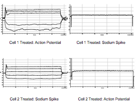
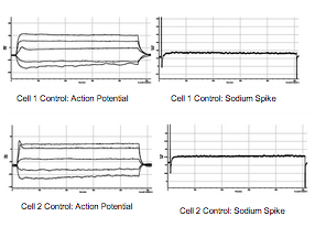
Little action potential or sodium spike was received from the untreated Neuroblastoma cells. A greater action potential and sodium spike was produced when testing the RA cells as shown in the graphs below.
|
